People with chronic kidney disease (CKD) commonly suffer from disorders of bone and mineral metabolism. Since nutritional imbalances are also common in CKD, it is likely one major contributing factor, among others. Optimizing vitamins and nutrients for bone health is therefore an important goal when managing people with CKD and bone disease. The interaction between CKD, vitamin D3 and bone disease is well established. In addition, there is growing evidence that vitamin K2 deficiency in CKD can influence vascular calcifications and bone health. But what about another bone health nutrient- vitamin A? Little is known about vitamin A and kidney health and the role of vitamin A in bone disease associated with CKD. This blog will discuss the role of vitamin A on bone health and explore its impact on bone disease in CKD.

By Majd Isreb, MD, FACP, FASN, IFMCP
What is vitamin A?
Vitamin A is an essential fat-soluble nutrient known for its role in good vision. It is also needed for cellular growth and differentiation. Since skin and gut cells are some of the fastest-growing cells in the human body, they are most sensitive to vitamin A deficiency. Vitamin A is also important for cells of the immune system. Vitamin A plays an important role in these processes:
- Growth and differentiation of all cells
- Embryonic development
- Organ formation in utero
- Normal immune function
- Eye development and vision
- Red blood cell production

Chemically, vitamin A is a group of organic compounds that share a beta-ionone ring with an isoprenoid chain. These compounds are often referred to as “retinoids.” The name of the retinoid depends on the number of the rings, the size of the isoprenoid chain, and the end group. These include -carotene, -carotene, retinal, retinol (which is vitamin A per se), all-trans-retinoic acid, all-trans-retinyl ester, 9-cis-retinoic acid, and 11-cis-retinal. Now, forget about all these different compounds, and let’s simplify things by calling them all vitamin A.
Diet and vitamin A
Vitamin A is consumed in the diet either as preformed vitamin A or as a precursor provitamin A carotenoid. Provitamin A carotenoids are converted to vitamin A in human intestinal cells. Sources of preformed vitamin A include eggs, liver, butter, milk, and fortified cereals. Provitamin A carotenoids are mainly found in vegetables such as carrots, spinach, collards, pumpkins, and squash. The most common type is beta-carotene. On average, the standard American diet provides approximately 700-800 micrograms of “retinol activity equivalents (RAE)” per day. Most of that comes as preformed vitamin A. The recommended dietary allowance of RAE for men and women is 900 and 700 micrograms/day, respectively. This makes deficiency of vitamin A rare. I will not discuss the complex absorption of vitamin A in the gut and storage in the liver and cellular mechanisms of action in detail here. These are described in detail elsewhere. However, it is important to remember that the presence of fat in the diet greatly enhances vitamin A absorption since it is fat-soluble. It is also worth noting that excessive intake of preformed vitamin A can cause elevated vitamin A levels. But elevated vitamin A does not usually occur after increased intake of provitamin A carotenoids. This is because the conversion of carotenoids to vitamin A is regulated by a negative feedback loop. When adequate levels of vitamin A are present, the body downregulates the production of vitamin A from carotenoids.
Join us to end the kidney disease epidemic
Effects of vitamin A on the bone
Before we discuss the effects of vitamin A on bone in detail, we should review the processes of bone formation and maintenance.
Bone formation and maintenance
Our bones contain a small number of cells surrounded by a mesh of collagen fibers that provide a scaffolding for salt crystals. These salt crystals are made of calcium, phosphate, and carbonate which combine to create, the so-called, hydroxyapatite. The latter incorporates other salts like magnesium hydroxide, fluoride, and sulfate as it hardens, or calcifies, on the collagen scaffolding. Hydroxyapatite crystals give bones their hardness and strength, while collagen fibers give them flexibility.
There are four types of cells that are found within the bone: osteoblasts, osteocytes, osteogenic cells, and osteoclasts. In adults, two processes are responsible for changes in the skeleton: modeling and remodeling.
Bone modeling describes the process of new bone formation or bone resorption on a given bone surface. This process is important for bone growth and shaping during childhood and adolescence. Bone remodeling, on the other hand, is the process that is used to maintain and renew healthy bones during adulthood. In other words, in bone modeling either bone formation or bone resorption occurs, while in bone remodeling both bone resorption and bone formation occur together.
For remodeling to occur the bone must be “dissolved,” and then a new bone is formed. In this process, osteoclasts dissolve old bone tissue at specific sites. This process is called resorption. Subsequently, new bone tissue is formed by the osteoblasts. Even though it may seem counterintuitive, bone resorption (breaking down old bone) is necessary for building new, healthy bone. So, in essence, modeling leads to the formation of new bone tissue where needed, while remodeling helps maintain and strengthen existing bone tissue.

The role of vitamin A
Vitamin A has two different effects on bone depending on the dose. While adequate vitamin A intake was shown to maintain healthy bones, high levels of vitamin A have been shown to cause the opposite. Let me explain.
Good effects
Studies have shown that vitamin A is important for bone resorption. This is essential for maintaining healthy bone by remodeling as we discussed. In fact, adequate intake of vitamin A has been found to improve bone mineral density and decrease the risk of fractures.
Not-so-good effects
In the early 1900s, researchers found more osteoclasts in the bones of animals with high vitamin A levels. Later animal studies showed that excess vitamin A led to the formation of bones with a “moth-eaten appearance.” It was also demonstrated that vitamin A stimulated bone resorption. Recently, it was noted that vitamin A can stimulate mineral release and bone degradation in mouse bones.
These effects were blocked by osteoclast inhibitors such as bisphosphonate and calcitonin. In essence, vitamin A can increase osteoclast formation and differentiation causing increased bone resorption. This is good for the maintenance of healthy bones but becomes harmful when there is excessive resorption at high levels of vitamin A. The evidence that supports this comes from studies that showed an increased risk of hip fractures in the lowest and highest vitamin A blood levels.
There are other studies that also showed that a high daily intake of vitamin A was associated with decreased bone mineral density. In the NHANES study in the US, for example, daily vitamin A intake of more than 3,000 micrograms was associated with an increased risk of hip fracture. However, there was no increased risk of fractures with high beta-carotene intake. This low-dose versus high-dose phenomenon has been seen with other nutrients and is described as a U-shaped hormetic response. At low doses, there are symptoms of nutritional deficiency but at high doses, there are symptoms of toxicity.

The interaction between vitamin A & vitamin D3
As we noted before, nutrients don’t work solo. Optimal bone health requires optimization of vitamin D, vitamin K2, calcium, phosphorus, and magnesium. There have been reports of vitamins D and A opposing each other. Some studies showed that vitamin D protects against vitamin A toxicity. On the other side, excess vitamin A was also shown to reduce the effects of vitamin D toxicity. In humans, high vitamin A intake was found to decrease the ability of vitamin D to enhance calcium absorption in the gut. Studies have shown that the negative effects of excess vitamin A on bone mineral density and fracture risk are amplified when accompanied by vitamin D deficiency.
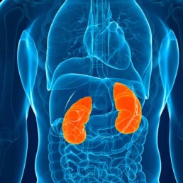
Vitamin A and kidney health
Several studies have shown that vitamin A levels increase with worsening kidney function and advanced CKD. This, along with the prevalence of vitamin D deficiency in this population, adds another layer of complexity to bone problems in kidney disease. This common complication of kidney disease is called chronic kidney disease-associated mineral bone disease (CKD-MBD).
In CKD-MBD, there is abnormal bone turnover and increased vascular and soft tissue calcifications. Increasing intake of preformed vitamin A at the advanced stage of CKD can lead to worsening bone abnormality and elevated calcium levels in the blood. This, in turn, can increase the risk of vascular calcifications. Therefore, supplementing vitamin A in patients with advanced CKD is not generally recommended on a regular basis. Natural vitamin A intake through a diet high in carotenoids should be sufficient.

Assessing vitamin A status
Unfortunately, it is difficult to assess vitamin A status in an individual. This is because most vitamin A is stored in the liver and is released as needed to the blood. The two common ways to measure vitamin A status are measuring serum retinol and retinyl ester concentrations. There are also labs that measure beta-carotene levels.
Serum retinol levels are only helpful if they are very low or very high. Levels < 1.05 micromol/L indicate vitamin A insufficiency. It has been suggested to use the ratio of serum retinyl esters to total serum vitamin A (retinol plus retinyl esters) as a marker for excess vitamin A. Serum retinyl ester levels exceeding 10% of total serum vitamin A may reflect excess vitamin A stores and potential toxicity.

How much vitamin A should I take?
Considering the above, we recommend our patients get most of their vitamin A from the diet (either as preformed vitamin A or provitamin A carotenoids). CKD patients can eat eggs, liver, butter, milk, carrots, spinach, collards, squash, and pumpkin with guidance from their dietitian/nutritionist and nephrologist. The following recommendations for CKD patients are based on anecdotal practice since the literature doesn’t support specific recommendations.
Patients with stage I-IIIa CKD may take up to 2500-3000 IU of supplemental vitamin A (or RAE 900 microgram/day for men and 700 microgram/day for women.) Patients with stage IIIb-IV CKD should decrease their intake of vitamin A supplements by 50%. Supplementation with vitamin A is not recommended for patients with an estimated GFR of less than 20 ml/min. If supplementation is desired during periods of sickness (for example, a respiratory illness) to boost the immune system, we recommend using beta-carotene supplements instead of preformed vitamin A supplements.
The Bottom Line
Vitamin A has a great impact on bone health. It is essential for bone resorption and remodeling. However, excess vitamin A intake can lead to bone weakness and fractures. In patients with advanced CKD, vitamin A tends to accumulate, and supplementation is not recommended. If supplementation is desired to boost the mucosal immune system during periods of sickness and high demand, look for supplements that provide much of their vitamin A in the form of beta-carotene. The individualized integrative approach to CKD-MBD necessitates careful nutritional assessment and evaluation of vitamin A levels in addition to vitamins D, K, magnesium, calcium, and phosphorus. This can help develop a tailored lifestyle and nutrition plan that can be incorporated into the medical management of CKD.