Polycystic kidney disease (PKD) is the most common inherited kidney disease affecting one in 800 people. It is passed down from parent to child over generations, through classic Mendelian genetics. There have been several advances in our understanding of PKD since our last blog. This blog will discuss the genetics of PKD. In the next blog we will discuss recommendations for water, salt, and caffeine intake to reduce cyst growth. There, you will find out if ketogenic diet or intermittent fasting is the best diet for polycystic kidney disease.

Hereditary Kidney Disease: The Genetics of PKD
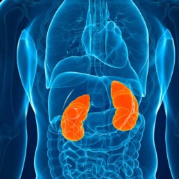
PKD is a group of two genetic disorders that affect the kidneys and cause the formation of multiple fluid-filled cysts of various sizes. As these cysts grow, they squeeze and destroy normal kidney tissue, eventually leading to loss of kidney function. PKD is an inherited disease, meaning it’s passed down genetically from parent to child, just like eye or hair color. Polycystic kidney disease can be autosomal-dominant (ADPKD) affecting 50% of offspring. Or it can be autosomal-recessive (ARPKD), affecting 25% of offspring.
Mutations in one of two genes (PKD1 or PKD2) account for most cases of ADPKD. Polycystic kidney disease 1 gene (PKD1) mutations are the most common. About 80 percent of patients affected by ADPKD have a PKD1 mutation. PKD2 gene is the cause of up to 20% of ADPKD cases and it is usually associated with milder course.
The PKD1 and PKD2 genes provide the blueprints for important kidney and liver proteins called polycystin-1 (PC1) and polycystin-2 (PC2). These proteins are crucial for the structure of the kidney’s tubular cells, which filter and clean the blood. PC1 and PC2 influence healthy growth and fluid secretion in these cells. However, in people with hereditary kidney disease, abnormal genetic blueprints lead to the production of abnormal proteins. When these kidney proteins don’t work properly, cysts accumulate and damage the kidney.
Cysts that form in polycystic kidney disease usually occur when the cells lining the tubules of the kidney start growing out of control (called proliferation). These outgrowths bulge and eventually separate into cysts. As the cysts grow, they transport fluid across their lining, forming a fluid-filled sack much like a balloon. So, when we think about PKD, we should remember two processes: cell proliferation (cell growth) and fluid secretion into the cysts.
Truncating vs. Non-truncating mutations
Sometimes a change in the DNA sequence of a gene results in the creation of an “early stop.” This early stop functions to end the translation of the gene into a protein in our cell factory. This produces a shortened or truncated protein. This type of variant can have serious functional consequences. They are called truncating mutations.
The relationship between genetic variants and prognosis in PKD is not completely understood. In a study that looked at the “renal survival” in 741 patients with ADPKD, PKD2 mutations were associated with approximately 20 years longer survival than PKD1 mutations. In addition, the type of PKD1 mutation, not its position, correlated strongly with renal survival. The median age at onset of kidney failure was 55 years for carriers of a truncating mutation and 67 years for carriers of a non-truncating mutation. This observation allows the integration of genic and allelic effects into a single scheme, which may have prognostic value.
This points to the importance of genetic testing even in a genetic disease with an obvious clinical presentation such as PKD. Identifying patients with truncating mutations may help selecting those who require more aggressive therapies.
PKD at the Cellular Level
To determine the best PKD diet and lifestyle recommendations, we must understand what is happening at the cellular level. In essence, abnormalities in PC1 or PC2 proteins will activate two pathways inside the cell. First, it activates the cAMP pathway which regulates fluid transport. Second, it changes the way cells make energy from sugar (called glycolysis). When PC1 or PC2 proteins don’t work well, they switch from aerobic (with oxygen) to anaerobic (without oxygen) glycolysis. This is similar to the shift that occurs in cancer cells.
While anaerobic glycolysis produces less energy from glucose, it is faster than aerobic glycolysis. This faster energy production allows the cells to grow faster. However, it also leads to a critical dependence on glucose.1
In the next blog we will discuss the 2021 update about diet and Lifestyle Treatments to Improve Polycystic Kidney Disease (PKD).
1 It is noteworthy that glycolysis is inhibited by a cellular messenger called AMP-activated protein kinase (AMPK). It is activated by another messenger called mTOR. Metformin activates AMPK.