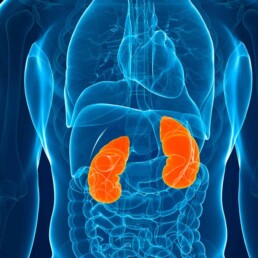
Each kidney contains several tiny filtering units called the nephrons. The nephron has two major sections: a filter, called the glomerulus (pleural: glomeruli), and the tubules. The tubules are divided into four sections. In this blog, we will focus mainly on the first section of the tubules, called the proximal tubules. We hope we will convince you that they are truly unique.

By Majd Isreb, MD, FACP, FASN, IFMCP
The proximal tubules
The proximal tubules are the site of retrieval of many electrolytes
The kidneys filter waste products from the blood in two stages. The first involves filtering a large amount of fluid through tiny blood vessels called glomeruli. In this first stage, 180-200 liters of blood are filtered daily! Considering that the total blood volume of the average adult is 4.5-5.7 liters, it is easy to realize that a person may die within a few hours if this was the only filtration step. Therefore, the second stage is so important. In it, millions of tiny tubes in the kidney, called tubules, reclaim most of that filtrate.
About 90% of this retrieval process occurs in the proximal tubule. The proximal tubules absorb 70% of the filtered sodium, 90% of the filtered bicarbonate, 60-70% of the filtered calcium, 70% of the filtered phosphate, and most of the filtered chloride. The proximal tubules also reclaim most of the filtered water (60%). In addition, up to 180 g/day of glucose is filtered by the glomerulus, and almost all of it is retrieved in the proximal tubule. The proximal tubules reabsorb all the filtered amino acids.
The proximal tubules are the site of many waste products and medication excretion.
Many small molecules are actively excreted in the proximal tubules. Drugs such as antibiotics, diuretics, nonsteroidal anti-inflammatory drugs, and others are excreted by the proximal tubules. Some of the gut-derived uremic toxins also get eliminated here. The proximal tubules are also the primary site for wasting metabolites such as folate, urate, creatinine, and carnitine. You can find a good list of these substances in this Wikipedia article.
Many specialized transporters are involved in this active transport. These include the families of organic anion transporters (OAT) and organic cationic transporters (OCT). Other transporters are also involved. It is, therefore, easy to understand that minor genetic changes in the genes that encode these transporters can lead to significant kidney diseases, including kidney stones.
Join us to end the kidney disease epidemic and receive the FREE Report “5 Pitfalls to Avoid When Caring for Kidney Patients”
The site of gluconeogenesis in the kidneys
The levels of blood sugar (glucose) levels are heavily regulated. Gluconeogenesis describes the process our body produces glucose from other precursors. This process is crucial for maintaining blood sugar balance in normal individuals. The liver is usually known to be the primary site for gluconeogenesis. However, recent data showed that the kidneys are also a significant site for gluconeogenesis.
After an overnight fast, the kidneys account for 40% of gluconeogenesis. Most of that is produced in the proximal tubules. The proximal tubules use lactate to produce glucose. This process is regulated by insulin, blood acid level, and stress hormones.
Cortisol appears to increase the production of glucose by the kidneys. Epinephrine (the fight or flight hormone) also can increase glucose release by the kidneys. This effect was eliminated by renal denervation. Insulin, on the other hand, decreases the release of glucose by the kidneys. In addition, elevated blood and urine glucose was found to reduce glucose production in the proximal tubules. Finally, high blood acid level has been found to increase glucose production by the kidneys.
The place of insulin degradation
Since we are talking about glucose balance, we should add that the kidneys are a significant site for insulin metabolism and clearance. The kidneys are responsible for 40% of insulin metabolism and clearance. In healthy individuals, the kidney clearance of insulin is 200 ml/min. This value is greater than GFR. This is because the glomeruli filter insulin. The proximal tubules then reabsorb it. Most of that gets broken down in the proximal tubular cells.
The proximal tubules are responsible for kidney clearance. This explains the insulin requirement decrease in diabetic patients who develop advanced kidney disease. It is also why many diabetic patients with progressive kidney disease develop low blood sugar while on glucose-lowering medications.
The site of vitamin D activation in the kidneys
Vitamin D is a fat-soluble vitamin essential for life and crucial for calcium balance and bone health. It is produced by our skin or consumed in the diet and then transported to the liver as a “prohormone” by a protein called Vitamin D Binding Protein (DBP). In the liver, this precursory form is converted to 25-hydroxyvitamin D (abbreviated 25(OH)D). 25-(OH)D is the primary circulating form of vitamin D. 25(OH)D is eventually transported to the kidneys, where another (-OH) group is added. The result is the formation of the active hormone called 1,25-dihydroxy vitamin D (or 1,25(OH)2D).
The kidneys not only convert 25(OH)D to the active hormone 1,25 (OH)2D, but they also reclaim 25(OH)D that is filtered in the first stage. The majority of this happens in the proximal tubules. In CKD, this process declines. This explains the high rate of vitamin D deficiency in CKD. In healthy individuals, when the proximal tubules reabsorb 25(OH)D, it gets either converted to 1,25(OH)2D or recycled back into the circulation.
Tubular secretion of creatinine
As we mentioned above, the proximal tubules secrete creatinine by OCT transporters. This secretion was found to increase with the worsening kidney function. Kidney function in clinical practice is measured by estimating the glomerular filtration rate (eGFR) based on serum creatinine levels. Since these levels are inaccurate due to increased tubular secretion of creatinine, studies have looked at another blood marker that is only filtered by glomeruli and not affected by the tubules. Cystatin C is one of these markers.
Testing tubular secretory function
Despite the importance of tubular secretory function, it is rarely measured or even estimated. The development of accurate and valuable tests of tubular secretion faces several challenges. The major challenge is finding a substance exclusively eliminated by the proximal tubules.
However, testing and measuring the proximal tubules’ secretory function is very important. It can:
- Predict the clearance of gut-derived uremic toxins.
- Allow early identification of specific kidney diseases
- Optimize medication dosing
- Estimate residual kidney function
- Predict metabolic complications of kidney disease
The bottom line
The proximal tubules are often underestimated and misunderstood in kidney health and disease. These sections of the kidneys are highly active and perform many biological functions. This explains why they utilize high energy and require significant nutritional and antioxidant support.